Deep Learning-Driven Automated Segmentation of High-Resolution 3D Histological Mouse Brain Volumes
Institutions
1. The National Institute of
Health and Medical Research (INSERM), France
2. ICMUB Laboratory,
CNRS UMR 6302, University of Burgundy, Dijon, France
3. NeuroGeMMLaboratory,
INSERM Unit
1231, University of Burgundy, Dijon, France
Introduction
Objective: Enhancing neurobiological studies through the development of an
automated segmentation
framework for high-resolution 3D histological mouse brain images.
Background: Importance of detailed and efficient brain imaging for
understanding neurological
development and disorders.
Methods
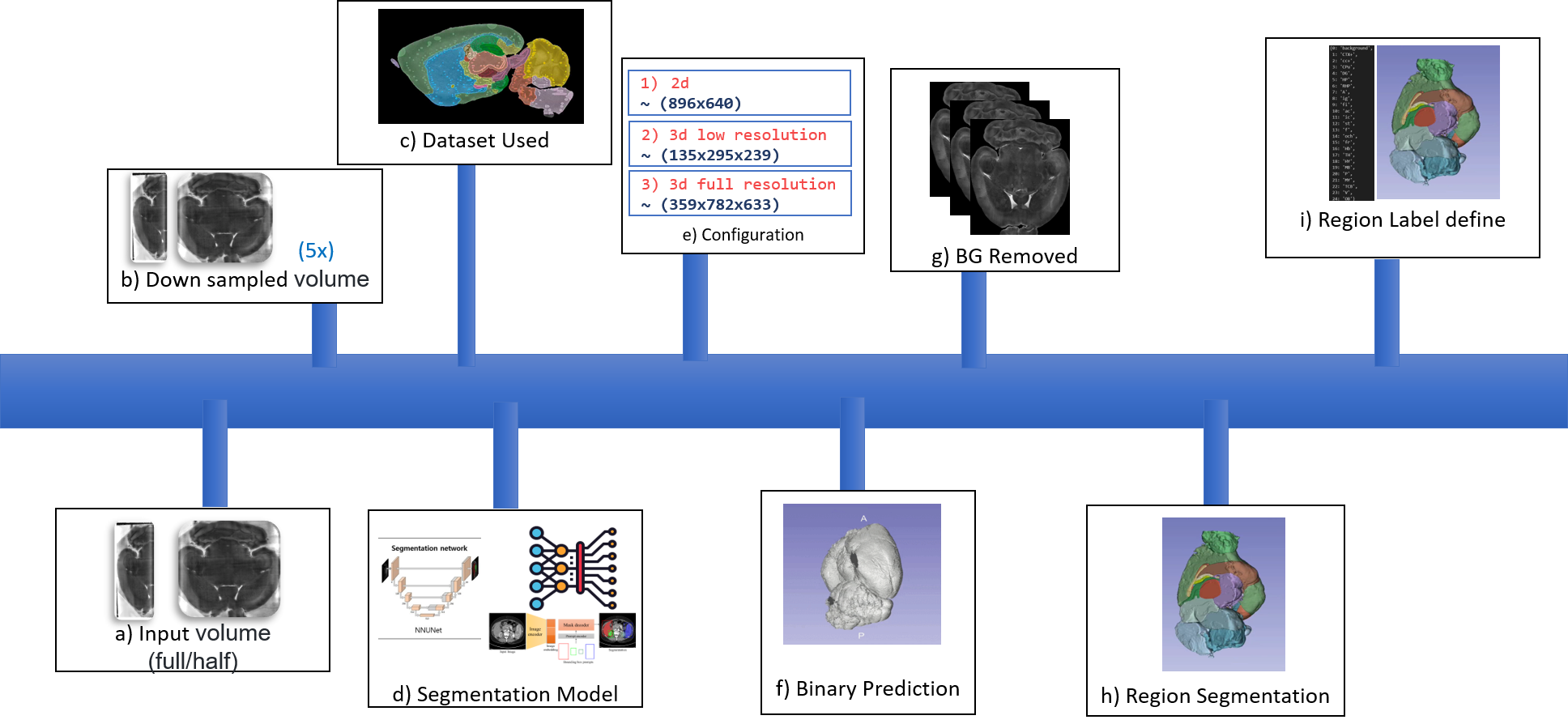
Deep Learning Models Used:
nnU-Net for automated pipeline configuration.
Segment Anything Model (SAM) adapted for 3D medical imaging.
Dataset: Private dataset consisting of nearly raw raster data (nrrd) format, with
volumes ranging
from 25 to 35 GB.
Results
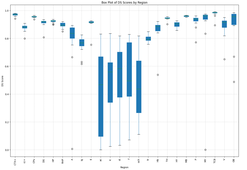
Performance Metrics:
Binary Segmentation DSC: 0.99
Multi-class Segmentation DSC: 0.87
Efficiency Gains: Reduced segmentation time from 30 hours to 5 minutes per
volume.
Visual Demonstrations:
Pipleline
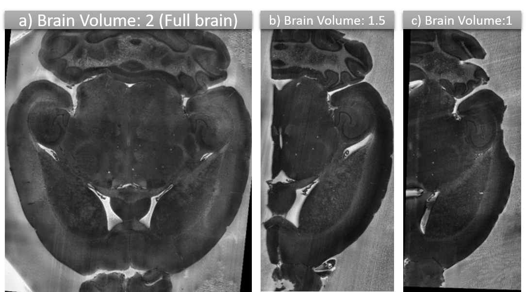
 Brain Volume Sequence
 Segmented Half and full brain

Segmented Half and full brain